Escondido Eyecare
  • Dr Verret
  • Contact Us
  • What to Expect
  • Promotions
  • Financing
​(760) 747-7979
Picture

Eye Anatomy

Picture
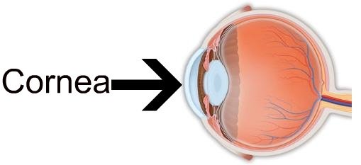
Cornea: Clear tissue in front of the color of the eye.
Picture
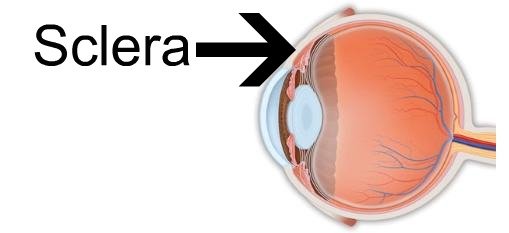
Sclera: White of the eye.
Picture
Conjunctiva: Clear tissue covering the sclera.
Picture
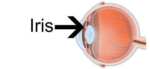
Iris: Color of the eye.
Pupil: Hole in the middle of the iris.
Aqueous: Liquid filling the space between the cornea and the iris.
Picture
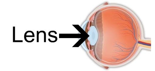
Lens: Part of the eye responsible for focusing.
Picture
Ciliary Muscle: Reshapes the lens to allow the eye to focus.
Picture
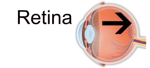
Retina: Composed of tiny receptors that allow the eye to see.
Vitreous: Clear gel filling the inside of the eye.
Picture
Macula: Center of the retina. Has the most densely packed receptors for best vision.
Picture
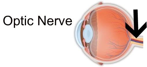
Optic Nerve: Connects the eye to the brain.
Picture
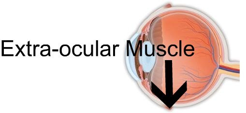
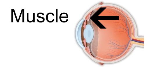
Extra-ocular Muscle: Allows the eye to move.
Picture
​(760) 747-7979
Picture
Home
Dr Eric Verret
Contact Us
Schedule Your Appointment
What to Expect
Promotions
Financing
Blog
Why Choose Us?
Meet our Team
About
Glamorous Glasses 
Fabulous Sunglasses
Safety Glasses and Sunglasses
​State-of-the-Art Contact Lenses
Lasik Consultation
Slowing Myopia Progression
Blue Light Glasses
Sunglasses Article
Dry Eye
Eye Strain
Definition of Optometry Terminology
Eye Anatomy
Guarantees and Warranties and Patient's Own Frame Waiver Policy
Notice of Privacy Practices
Terms of Service
Privacy Policy
Assignment of Benefits and Financial Agreement
Escondido Eyecare's Donations to Charities
Physician Patient Arbitration Agreement
Online Forms
​Site Map
​Copyright © 2022
  • Dr Verret
  • Contact Us
  • What to Expect
  • Promotions
  • Financing